Clinical Case Overview
Spinal deformity is a complex three-dimensional condition that may involve abnormal alignment in the coronal, sagittal, and axial planes. In patients with significant deformity, surgical correction may require a combination of deformity assessment, three-dimensional imaging, pedicle screw fixation, rod-based correction, and spinal fusion.
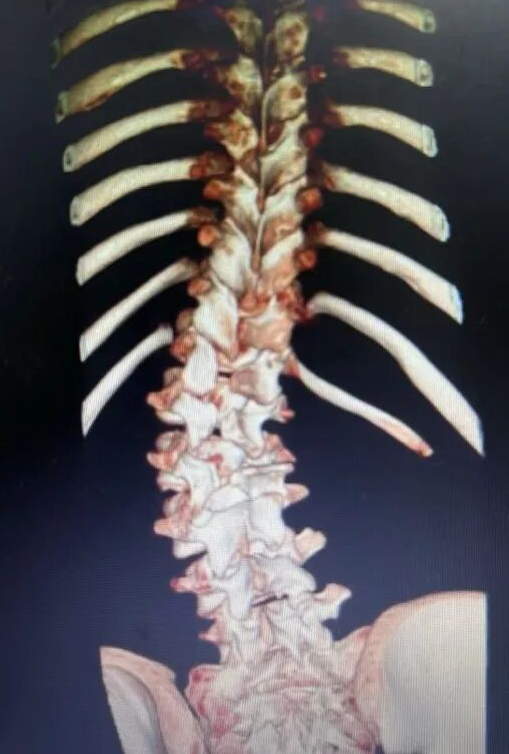
This article presents an authorized clinical imaging case of spinal deformity correction, including preoperative X-ray, CT and MRI evaluation and postoperative radiographic assessment.
The case demonstrates how detailed imaging evaluation can support surgical planning and how a posterior thoracolumbar rod-screw system can be incorporated into a comprehensive spinal deformity correction strategy.
| 3D Reconstructed Image |
 |
1. Clinical Presentation and Initial Imaging
The patient presented with a significant spinal deformity requiring comprehensive imaging evaluation.
The initial full-spine radiograph demonstrated abnormal spinal alignment and curvature. Because spinal deformity is a three-dimensional condition, plain radiographs alone may not provide sufficient information for detailed surgical planning.
Further imaging with CT and MRI was therefore performed as part of the preoperative assessment.
The imaging evaluation focused on:
The relationship between the deformity and surrounding anatomical structures
This multimodal imaging approach provided complementary information for understanding the deformity and planning posterior instrumentation.
| X-ray Image |
 |
2. CT and MRI Evaluation
CT Imaging
CT imaging provided more detailed information about the vertebral anatomy and bony structures.
For complex spinal deformity cases, CT may assist in evaluating:
This information can be particularly important when planning the placement of pedicle screws.
MRI Evaluation
MRI provides complementary information about the spinal canal, spinal cord, nerve roots, intervertebral discs and other soft-tissue structures.
In a spinal deformity case, MRI findings can help identify associated neural or soft-tissue abnormalities that may influence surgical planning.
| Magnetic Resonance Image |
 |
3. Preoperative Assessment and Surgical Planning
The combination of X-ray, CT and MRI allowed a more comprehensive assessment of the spinal deformity.
For posterior spinal deformity surgery, implant planning may include consideration of:
The exact surgical plan should be individualized according to the patient's anatomy, deformity characteristics and clinical condition.
For complex deformities, segmental pedicle screw fixation can provide multiple points of fixation along the spine and facilitate controlled correction using a rod-screw construct.
4. Posterior Rod-Screw Fixation for Spinal Deformity Correction
The posterior rod-screw construct is an important component of modern spinal deformity surgery.
A typical posterior thoracolumbar fixation construct may include:
ATOM's Ti Posterior Thoracolumbar Rod-Screw System (5.5) is a comprehensive posterior fixation system incorporating pedicle screws, titanium rods, locking components, connectors and other auxiliary components. The system is designed for posterior fixation of the thoracic, lumbar, lumbosacral and iliac regions and includes applications such as spinal deformity and scoliosis.

5. ATOM Ti Posterior Rod-Screw for Spinal Deformity (5.5 System)
For this clinical case, the article can naturally introduce ATOM's dedicated Ti Posterior Rod-Screw for Spinal Deformity (5.5 System) as the product solution associated with the deformity correction concept.
ATOM identifies the product as:
Product Name: Ti Posterior Rod-Screw for Spinal Deformity (5.5 System)
Model Number: 5.5/6.0
Application: Spinal Deformity
Compatible Instrument: ACB03
The product is designed to work with compatible 5.5/6.0/6.35 components for vertebral rotation correction during spinal deformity correction procedures.

6. Vertebral Rotation Correction
One of the important considerations in spinal deformity surgery is that correction is not limited to the coronal plane.
Scoliosis and other complex spinal deformities may involve:
Coronal deformity + sagittal imbalance + axial vertebral rotation
Therefore, a comprehensive correction strategy may need to address multiple planes of deformity.
ATOM's dedicated spinal deformity correction component is designed to support vertebral rotation correction and multiple vertebral linkage rotation correction.
The product design incorporates a triangular structure intended to engage the vertebral construct during rotation correction and distribute the applied forces.
7. Multi-Level Linked Rotation Correction
Complex spinal deformities frequently involve multiple vertebral levels rather than a single isolated vertebra.
ATOM's spinal deformity correction component is designed to support multiple vertebral linkage rotation correction, allowing correction forces to be coordinated across multiple instrumented levels.
This concept is particularly relevant when the surgical objective includes correction of rotational deformity across a multi-level construct.
Recommended SEO keywords in this section
Naturally incorporate:
multi-level spinal deformity correction
vertebral rotation correction
scoliosis correction
posterior rod-screw system
pedicle screw fixation
three-dimensional spinal deformity correction
Do not repeatedly insert the exact keyword in every paragraph. One or two natural occurrences per section are sufficient.
8. Flexible Rod Placement During Correction
Another product feature described by ATOM is that the rod can be placed before, during or after rotation correction, depending on the surgical technique and correction sequence.
This design is intended to provide flexibility during intraoperative correction.
The exact correction sequence should, however, be determined by the surgical technique, deformity characteristics, implant configuration and surgeon judgment.
9. Screw Options for Posterior Spinal Fixation
A posterior thoracolumbar rod-screw system may require different screw configurations depending on the anatomical level and correction strategy.
ATOM's 5.5 system includes several screw options, including:
Polyaxial screws
Reduction polyaxial screws
Monoaxial screws
Reduction monoaxial screws
Other compatible fixation components
ATOM's polyaxial screws are manufactured from titanium alloy TC4 and are listed for posterior internal fixation of the thoracic, lumbar, lumbosacral, coccygeal and iliac regions.
10. Postoperative Imaging
The postoperative radiographs in this case provide an important opportunity to evaluate the final spinal alignment and instrumentation.
Compared with the preoperative images, the postoperative X-rays should be used to demonstrate:
| Postoperative Images |
 |
| Comparison of images of patients before and after surgery |

|
Conclusion
This authorized clinical case demonstrates the importance of comprehensive imaging and individualized surgical planning in complex spinal deformity correction.
Preoperative X-ray, CT and MRI provide complementary information for evaluating spinal alignment, vertebral anatomy and relevant neural structures. During posterior deformity correction, pedicle screw and rod constructs can provide segmental fixation and facilitate controlled correction maneuvers.
The ATOM Ti Posterior Rod-Screw for Spinal Deformity (5.5 System) is specifically designed for spinal deformity applications and incorporates features intended to support vertebral rotation correction, multi-level linked rotation correction and flexible rod placement during correction.
Together with the broader Ti Posterior Thoracolumbar Rod-Screw System (5.5), compatible pedicle screws and dedicated spinal deformity instruments, the system forms part of ATOM's portfolio for posterior spinal fixation and deformity correction.